Case of the month
The Case of the Month aims to share interesting and educational genito-urinary pathology cases with the ISUP membership. All ISUP members are cordially invited to submit cases to the case manager (cotm@isupweb.org) using the available template (word document). Your submission is highly appreciated!
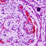
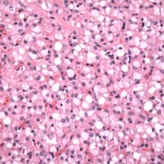
January 2017
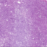
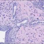
44-year-old man with right renal mass, retroperitoneal and mediastinal lymphadenopathy
An adrenal-sparing left radical nephrectomy was performed
Patient underwent several cycles of chemotherapy and biological treatment
Patient is alive with residual disease (retroperitoneal lymph nodes) 9 years after nephrectomy
December 2016
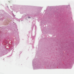
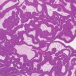
71 year-old male with a 3.4 cm, mid-pole, left renal mass
An adrenal-sparing left radical nephrectomy was performed
Gross Description
Radical nephrectomy specimen
Single 3.4 cm yellow-brown, focally hemorrhagic mass in the mid-pole aspect of the kidney
Tumour grossly confined to the kidney – abutting the middle renal calyx and bulging into the renal sinus
No involvement of the renal vein or its segmental branches
November 2016
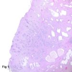
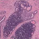
A 68 year-old male with HIV and clinical benign prostatic hyperplasia underwent TURP.
In one of the TURP chips a glandular proliferation is seen streaming between benign glands.
October 2016
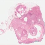
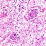
A 53 year-old female with an incidentally found 2.9 cm right renal mass on abdominal ultrasound.
Percutaneous needle biopsy was performed
limited lesion sampling (1-2 mm of tumor in a 15 mm core)
bland ovoid and spindle cells with no overt malignant features
positive for CD34 and SMA; negative for epithelial markers
reported as “mesenchymal neoplasm – definitive classification not possible on biopsy”
Patient chose surgical management over active surveillance with serial imaging
A right partial nephrectomy was performed
JULY 2016
– 55 year old male with long term use of exogenous testosterone.
– PSA- 6.9 ng/ml
– Digital rectal exam – large, firm prostate gland
– U/S imaging – anterior nodule, possible extraprostatic disease
– TRUS guided biopsies – 12 core set, anterior nodule not targeted for biopsy
June 2016
64 year old male
5 year history of intermittent gross hematuria
Social history: Non-smoker, metal plater with exposure to trichloroethylene 15-20 years ago
No history of previous bladder lesions
3.5 cm bladder neck / trigone tumor
Cystoscopic examination: Papillary tumor with a narrow stalk, suspicious for urothelial carcinoma
Treatment: Transurethral resection of tumor
May 2016
– 35 year old female with a history of ovarian serous borderline tumor (treated by resection only)
– Incidentally detected heterogeneous, enhancing 5 cm upper pole renal mass on an otherwise negative follow-up CT scan for her ovarian tumor
– Lesion was deemed suspicious for renal cell carcinoma (RCC) by imaging criteria
– Right radical nephrectomy was recommended and performed, with a 6.3 cm predominantly solid mass in the upper pole of the right kidney
APRIL 2016
64 year-old male
PSA – 33 ng/ml
Digital rectal exam – right posterolateral nodule
Prostate biopsy – Gleason 8/10 (4+4) (ISUP 4), involving 1 of 12 cores and 100% of the positive core, right lateral
Staging work-up – negative for metastases
Treatment – radical prostatectomy performed according to patient preference
MARCH 2016
57 year old man presented with a 3 cm incidental renal mass. A tumorectomy was performed. Patient is alive and well 7 months after surgery
February 2016
Clinical history: 57 y/o male with 3 cm right renal mass. Partial nephrectomy was performed