Case of the month
The Case of the Month aims to share interesting and educational genito-urinary pathology cases with the ISUP membership. All ISUP members are cordially invited to submit cases to the case manager (cotm@isupweb.org) using the available template (word document). Your submission is highly appreciated!
August 2017
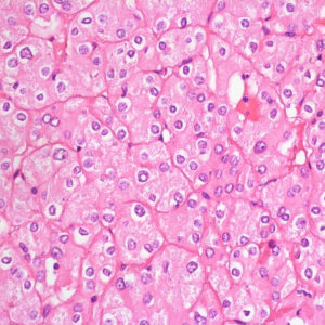
61 year-old man with renal mass
July 2017
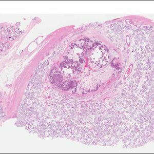
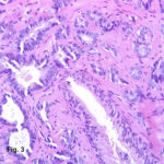
A 63-year-old male with prostate needle biopsy
June 2017
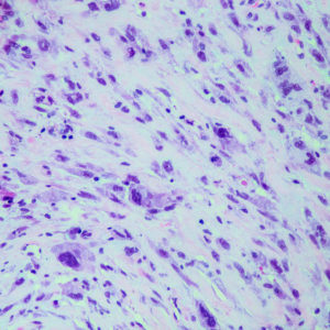
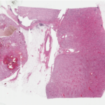
49 year-old male who presented with a right inguinal hernia and right testicular mass
May 2017
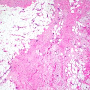
A 54 year old male with past medical history of hydrocele, presents with onset of bilateral scrotal masses and buried penis 6 months status post uncomplicated left inguinal herniorrhaphy. Sonographic imaging demonstrated skin thinking with underlying amorphous subcutaneous mass involving the scrotum, left > right, with compression of and atrophy of the left testis. Patient underwent resection of bilateral scrotal masses, which measured 21×16.5×4.5cm and 14x9x3.5cm.
April 2017
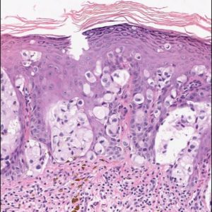
81 year-old man presenting with an isolated leukoplakia-like penile (preputial) lesion of unknown duration.
A 0.3 cm incisional biopsy was performed.
March 2017
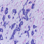
A 73-year- old male with LUTS and s-PSA 6.6. A 10-core prostate biopsy was taken. In addition to a conventional prostate cancer GS 7 (neg for p63, pos for AMACR), a glandular proliferation is seen in 4 cores.
February 2017
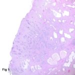
A 42-year-old male with right ureteric stricture
4-year history of recurrent right ureteric calculi, status post ureteroscopic lithotripsy (USRL) twice
Cystoscopy: Stenosis of right ureteric orifice, which was unable to pass guide wire
Treatment: Ureteric reimplantation with lysis of adhesion was performed; ureteric wall was sent to pathology
Gross findings: Thickened ureteric wall lined by smooth and flat mucosa
January 2017
44-year-old man with right renal mass, retroperitoneal and mediastinal lymphadenopathy
An adrenal-sparing left radical nephrectomy was performed
Patient underwent several cycles of chemotherapy and biological treatment
Patient is alive with residual disease (retroperitoneal lymph nodes) 9 years after nephrectomy
December 2016
71 year-old male with a 3.4 cm, mid-pole, left renal mass
An adrenal-sparing left radical nephrectomy was performed
Gross Description
Radical nephrectomy specimen
Single 3.4 cm yellow-brown, focally hemorrhagic mass in the mid-pole aspect of the kidney
Tumour grossly confined to the kidney – abutting the middle renal calyx and bulging into the renal sinus
No involvement of the renal vein or its segmental branches
November 2016
A 68 year-old male with HIV and clinical benign prostatic hyperplasia underwent TURP.
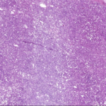
In one of the TURP chips a glandular proliferation is seen streaming between benign glands.