Case ID: 1198
Publication date: 15 Jan, 2025
Consensus grade: Other
Show diagnosis by expert panel members| User | Diagnosis | Difficulty | Comment |
|---|---|---|---|
| Pathologist 1 | Other | Typical |
This appears to be an epithelioid angiomyolipoma |
| Pathologist 2 | Other | Not typical |
?AML variant, ?sarcomatoid RCC ? met. Is there pigment in the cells? Technically sub-optimal images. |
| Pathologist 3 | Other | Not typical |
RCC vs angiomyolipoma |
| Pathologist 4 | Other | Not typical |
Epithelioid PEComa |
| Pathologist 5 | Other | Typical |
Epithelioid AML |
| Pathologist 6 | Other | Not typical | |
| Pathologist 7 | Insufficient tumor for diagnosis | Not typical |
morphology bad |
| Pathologist 8 | Renal cell carcinoma, unclassified | Not typical | |
| Pathologist 9 | Other | Not typical |
Terrible images |
| Pathologist 10 | Other | Typical | |
| Pathologist 11 | Other | Typical |
Based on these images alone, would not call pure epithelioid AML |
| Pathologist 12 | Other | Typical | |
| Pathologist 13 | ALK arrangement-associated RCC | Typical | |
| Pathologist 14 | Other | Typical | |
| Pathologist 15 | Other | Not typical |
AML/PEComa? |
| Pathologist 16 | Other | Not typical | |
| Pathologist 17 | Other | Not typical |
could be a number of diagnoses, epi AML, translocation RCC etc |
| Pathologist 18 | Clear cell RCC | Typical | |
| Pathologist 19 | Clear cell RCC | Not typical |
WHO/ISUP grade 4. Sarcomatoid/rhabdoid |
| Pathologist 20 | Other | Not typical | |
| Pathologist 21 | Other | Not typical |
Epithelioid angiomyolipoma? |
| Pathologist 22 | Clear cell RCC | Not typical |
Hopefully clear cell RCC with giant cells :-) |
Case description (by case creator):
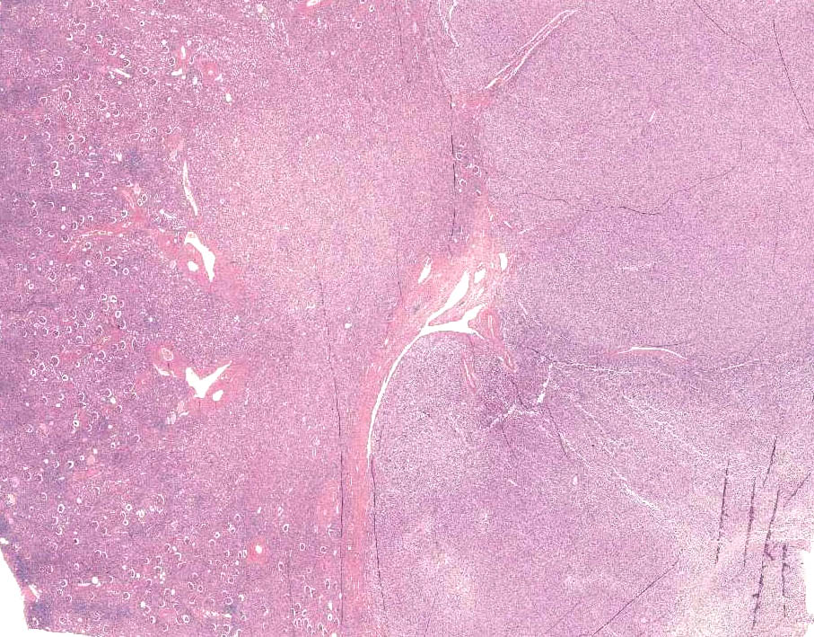
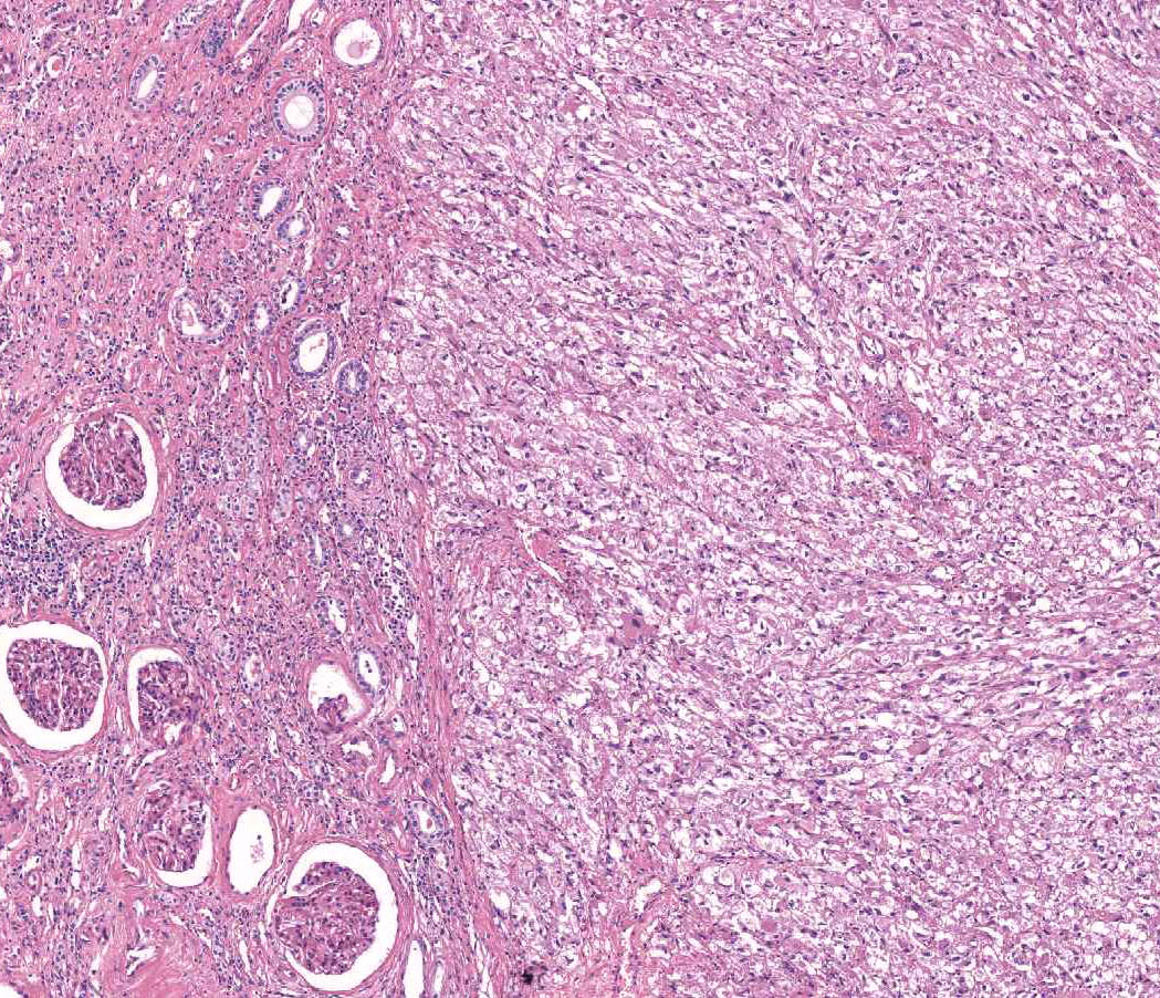
Mesorenal mass of 6 cm on the right kidney. Radical nephrectomy.
Cut surfaces: homogeneus and tan with large, fresh hemorrhagic areas.
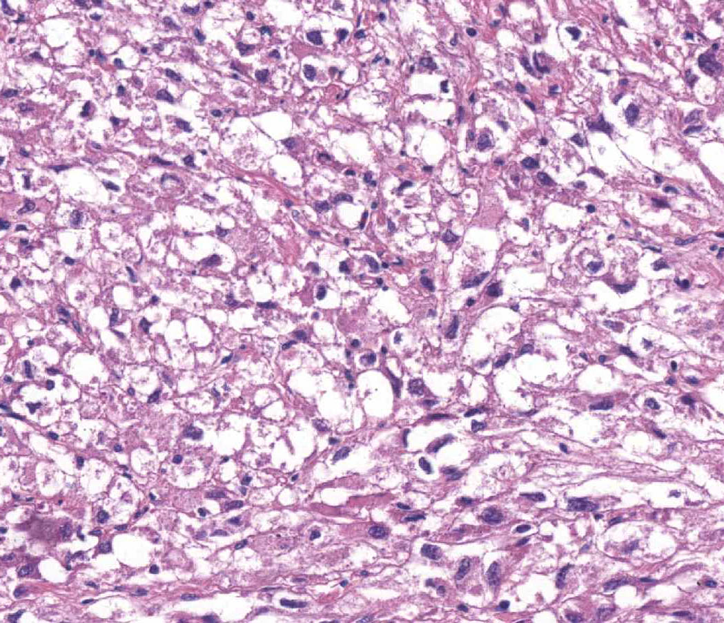
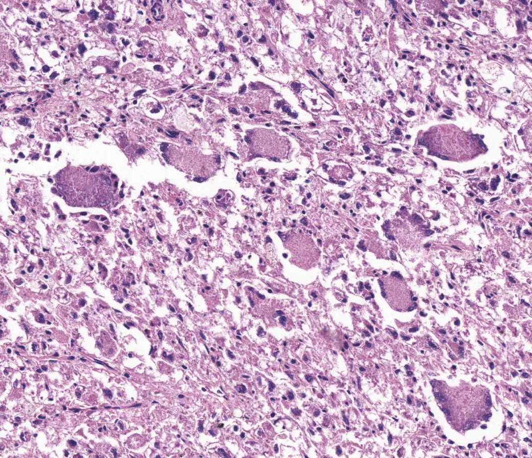
Microscopic examination: tumour with diffuse architecture, composed of neoplastic cells with epithelioid appearance and clear or granular cytoplasm.


