Case ID: 1121
Publication date: 29 Jan, 2025
Consensus grade: Succinate dehydrogenase deficient renal carcinoma
Show diagnosis by expert panel members| User | Diagnosis | Difficulty | Comment |
|---|---|---|---|
| Pathologist 1 | Succinate dehydrogenase deficient renal carcinoma | Typical | |
| Pathologist 2 | Succinate dehydrogenase deficient renal carcinoma | Not typical | |
| Pathologist 3 | Succinate dehydrogenase deficient renal carcinoma | Typical | |
| Pathologist 4 | Succinate dehydrogenase deficient renal carcinoma | Not typical |
Needs SDH staining to confirm. TSC-associated RCC is in the differential. |
| Pathologist 5 | Succinate dehydrogenase deficient renal carcinoma | Typical | |
| Pathologist 6 | Succinate dehydrogenase deficient renal carcinoma | Typical | |
| Pathologist 7 | Succinate dehydrogenase deficient renal carcinoma | Typical | |
| Pathologist 8 | Succinate dehydrogenase deficient renal carcinoma | Typical | |
| Pathologist 9 | Other | Not typical | |
| Pathologist 10 | Succinate dehydrogenase deficient renal carcinoma | Typical | |
| Pathologist 11 | Succinate dehydrogenase deficient renal carcinoma | Typical | |
| Pathologist 12 | Succinate dehydrogenase deficient renal carcinoma | Typical | |
| Pathologist 13 | Succinate dehydrogenase deficient renal carcinoma | Typical | |
| Pathologist 14 | Succinate dehydrogenase deficient renal carcinoma | Typical | |
| Pathologist 15 | Succinate dehydrogenase deficient renal carcinoma | Typical | |
| Pathologist 16 | Succinate dehydrogenase deficient renal carcinoma | Typical |
I would support my dg by IHC and/or analysis of SDH genes |
| Pathologist 17 | Clear cell RCC | Not typical | |
| Pathologist 18 | Succinate dehydrogenase deficient renal carcinoma | Typical | |
| Pathologist 19 | oncocytoma | Typical |
Case description (by case creator):
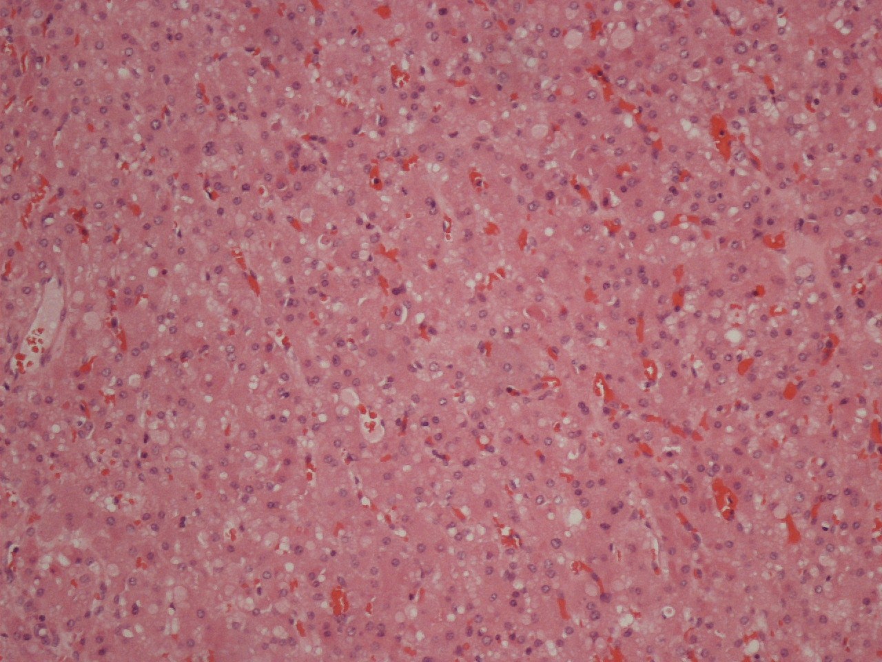
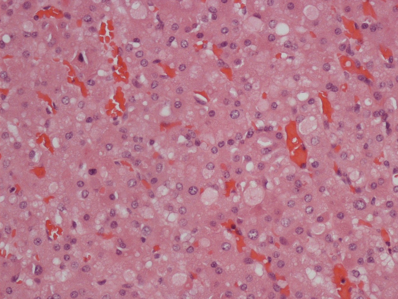
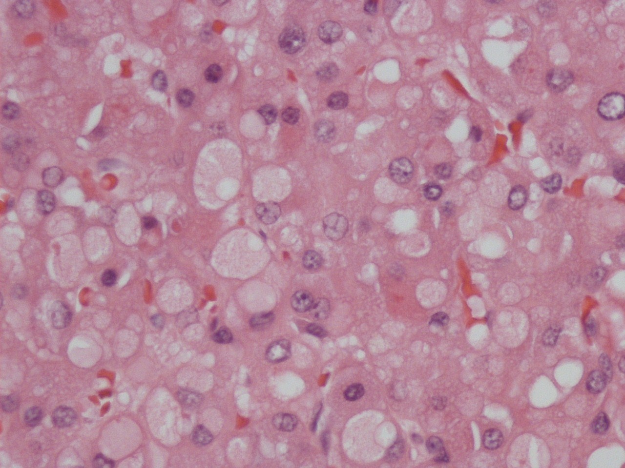
Renal tumour 42 year old woman. The tumor was 6cms in diameter with a homogeneous light tan color. The tumor has a solid architecture composed of trabeculae or tubules supported by a rich capillary network. The tumor cells are cuboidal with granular eosinophilic cytoplasm. Many cells contain intracytoplasmic inclusions which are round, appear membrane bound with a pale interior. These inclusions are large and cause lateral nuclear displacement. Nuclei are round exhibiting mild pleomorphism and nucleoli visible with a high power objective. Mitoses are infre quent.Necrosis and hemorrhage are not seen.