Case ID: 298
Publication date: 27 Apr, 2024
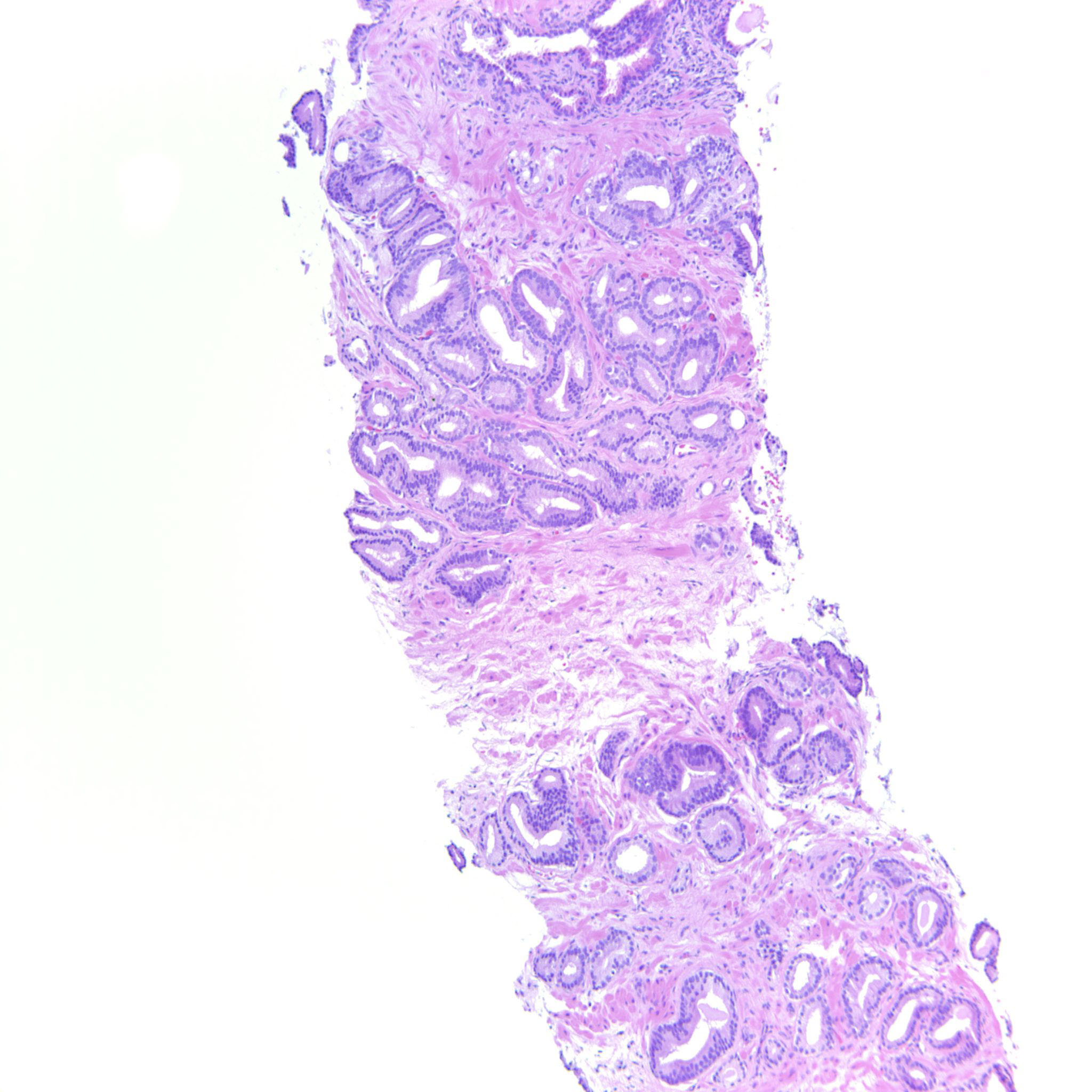
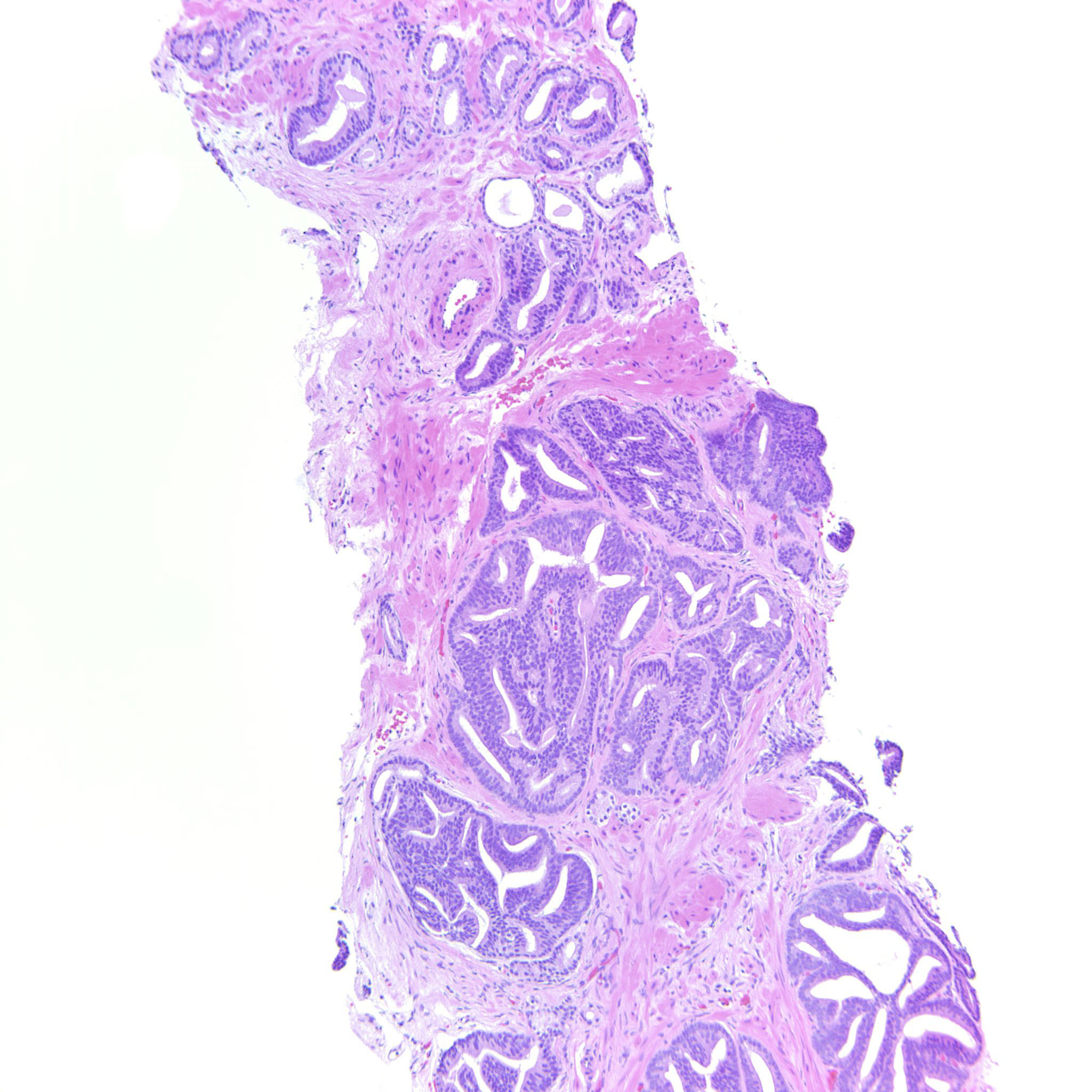
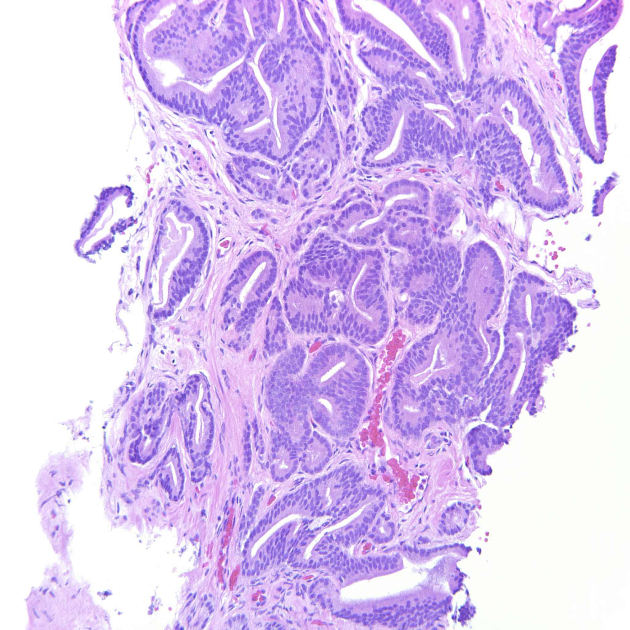
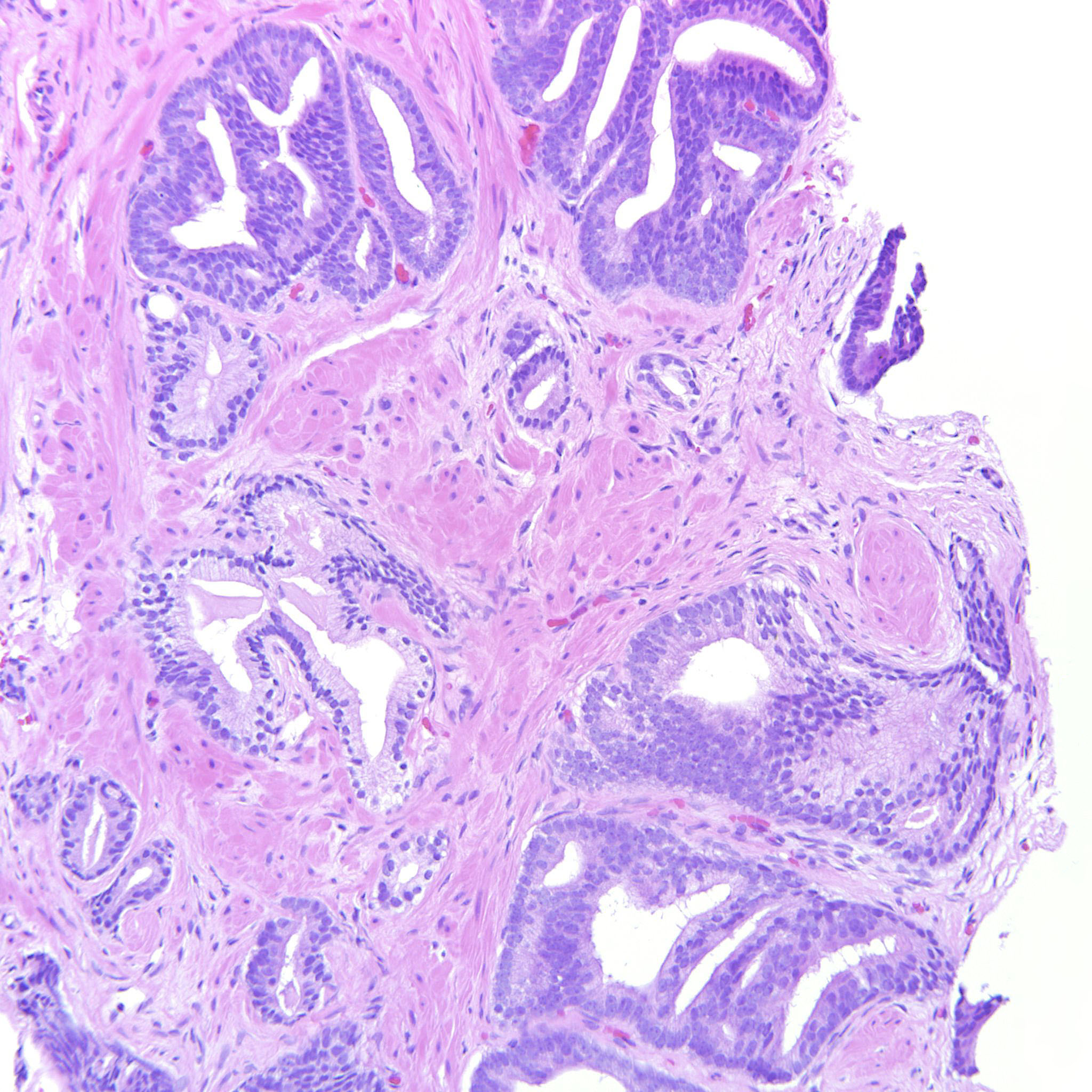
Consensus grade: GS 3+4=7 (ISUP 2)
Show diagnosis by expert panel members| User | Diagnosis | Difficulty | Comment |
|---|---|---|---|
| Pathologist 1 | GS 3+4=7 (ISUP 2) | Typical | |
| Pathologist 2 | GS 3+4=7 (ISUP 2) | Typical | |
| Pathologist 3 | GS 3+4=7 (ISUP 2) | Borderline higher |
Either ductal or IDC component |
| Pathologist 4 | GS 3+4=7 (ISUP 2) | Typical |
Find it difficult distinguishing 3+4 from 4+3 on separate images as some same area at different magnification. |
| Pathologist 5 | GS 3+4=7 (ISUP 2) | Borderline higher |
need better low power image |
| Pathologist 6 | GS 3+4=7 (ISUP 2) | Typical |
difficult to say whether "+4 or 4+3, because I cannot estimate which part is predominant on the pics. But 4 has to be in as well as 3 |
| Pathologist 7 | GS 4+3=7 (ISUP 3) | Typical |
Nice classic histologic patterns, but hard to tell relative percentage of each component |
| Pathologist 8 | GS 3+4=7 (ISUP 2) | Typical |
Should be straightformard but close to 50:50 3:4. |
| Pathologist 9 | GS 3+4=7 (ISUP 2) | Typical | |
| Pathologist 10 | GS 3+4=7 (ISUP 2) | Borderline higher |
Depends on the overall amount of 4 - low power overview image of the entire core would likely create greater agreement on 4+3 vs 3+4 |
| Pathologist 11 | GS 3+4=7 (ISUP 2) | Typical | |
| Pathologist 12 | GS 4+3=7 (ISUP 3) | Typical | |
| Pathologist 13 | GS 3+4=7 (ISUP 2) | Typical | |
| Pathologist 14 | GS 3+4=7 (ISUP 2) | Borderline higher |
difficult to estimate the amount of 3 and 4 by images; could be 4+3 |
| Pathologist 15 | GS 3+4=7 (ISUP 2) | Typical | |
| Pathologist 16 | GS 4+3=7 (ISUP 3) | Borderline lower |
Since photo 1 is the same as 6 iand the others are different, is difficult to gauge the amounts of 3 and 4. If all are considered different fields of 1 slide them cribriform 4 predominates |
| Pathologist 17 | GS 3+4=7 (ISUP 2) | Borderline lower |
is that cribriform area intraductal - does that make it 3+3 then!! |
| Pathologist 18 | GS 4+3=7 (ISUP 3) | Typical | |
| Pathologist 19 | GS 3+4=7 (ISUP 2) | Borderline higher |
Definitely both patterns 4 and 3 but it's tricky to estimate the relative percentages off the screen. I thinks it's about 40% pattern 4 and have graded it accordingly but can see others estimating the pattern 4 slightly higher. Also considered IDC-P and HGPIN focally and might have done a 34betaE12/p63 stain, depending on what the rest of the case looked like. Not convinced that there is a ductal adenocarcinoma component. |
| Pathologist 20 | GS 3+4=7 (ISUP 2) | Typical | |
| Pathologist 21 | GS 3+4=7 (ISUP 2) | Typical | |
| Pathologist 22 | GS 3+4=7 (ISUP 2) | Typical | |
| Pathologist 23 | GS 4+3=7 (ISUP 3) | Typical | |
| Pathologist 24 | GS 3+4=7 (ISUP 2) | Typical |
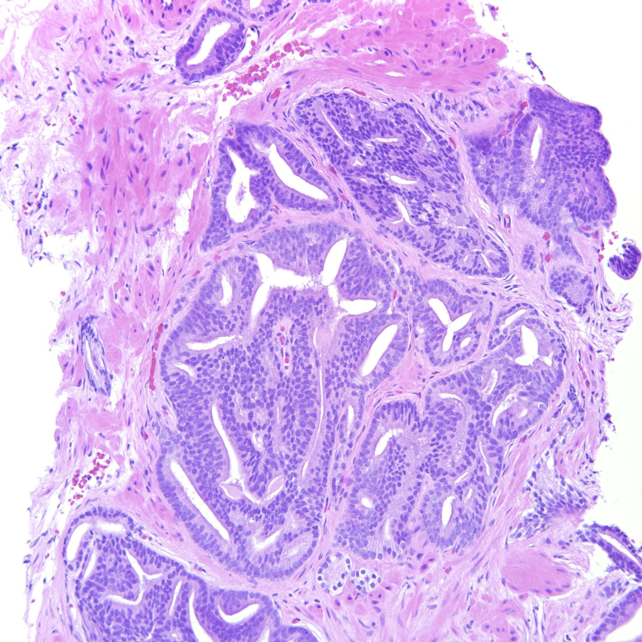
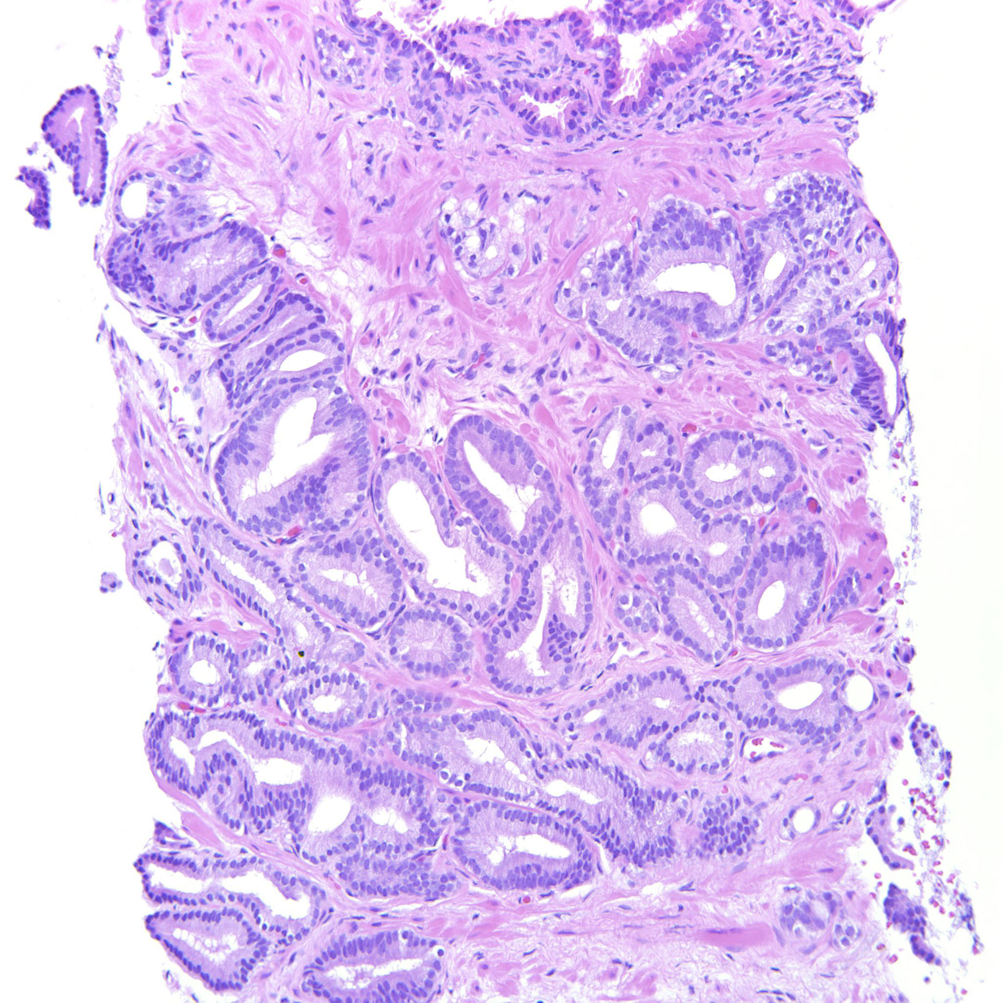
Case description (by case creator):
A lot of the tumor is GP3 but there are also a couple of cribriform glands. The epithelium is tall columnar, sometimes with elongated nuclei but the tumor does not have other features of ductal adenocarcinoma and should probably be considered a variant of acinar adenocarcinoma of the prostate.